Understanding A Tendon Injury
Conditions
Achilles Tendon Rupture
Bicep Tendon Rupture
Hamstring Tendon Rupture
Patellar Tendon Rupture
Quadriceps Tendon Rupture
Achilles Tendon Rupture
What is an Achilles Tendon Rupture?
The Achilles tendon is a group of tough fibrous tissue that connects the calf muscles to the heel bone. It functions so as to elevate the heel while walking or running. Overloading of this ligament can result in partial or full tearing of this tendon. Dr. Gregory DiFelice specializes in diagnosing and treating Achilles tendon ruptures.
Causes of Achilles Tendon Ruptures
Constant overuse or repetitive activities can cause Achilles tendon disorders. These activities exert excessive stress on the tendon and lead to micro tears. This damage or injury of the tendon results in pain. Reoccurring micro trauma can lead to Achilles tendinitis (inflammation) or tendinosis (degeneration).
People involved in activities like sports and exercises are more prone to develop Achilles tendon disorders. It is also commonly seen in people whose occupation puts lot of pressure on their feet and ankles. Simple movements like running, jumping, stretching, and improper shoes can also result in a complete rupture of the tendon.
Symptoms of Achilles Tendon Rupture
Symptoms related to Achilles tendinitis or tendinosis include swelling, mild pain, stiffness, and loss of range of motion. Patients who sustain a complete rupture of the Achilles tendon will report sudden pain and a pop, stating that they feel like they have been kicked in the back of the heel. Over the first 24, you may experience swelling and bruising are quite common, making it difficult to walk.
Diagnosis of Achilles Tendon Ruptures
To identify an Achilles tendon tear, Dr. Gregory DiFelice will ask about your medical history and perform a physical examination of your ankle. Some imaging tests, such as an X-ray or MRI scans, may be ordered to confirm the diagnosis. Dr. Gregory DiFelice will perform a Thompson squeeze test, by squeezing your calf. Patients with a complete tendon rupture will have minimal movement in their ankle.
Treatment of Achilles Tendon Ruptures
Achilles tendon tear can be treated by non-surgical and surgical methods based off the degree of injury. Partial tears to the Achilles will include non-surgical treatment, such as rest, ice, compression, and elevation (RICE protocol). A walking may be worn to help immobilize your ankle. Use of crutches may be recommended to protect your ankle. Physical therapy exercises may be recommended to improve ankle motion and strength. If you have a complete tear of the Achilles tendon, surgery will be recommended.
Surgical options for a complete tendon rupture include Achilles tendon repair. To perform this procedure, Dr. Gregory DiFelice will make an incision on the back of your ankle. The two ends of the tendon will then be surgically stitched together. You will then be placed into a walking boot immediately after surgery and instructed to walk with crutches. Following an Achilles tendon repair a rehabilitation program will be started that helps you resume a wider range of activities. Usually, a complete recovery may take about 6 to 12 months depending on your injury pattern.
For more information, please contact our office to schedule your appointment today.
Bicep Tendon Rupture
What is a Biceps Tendon Rupture?
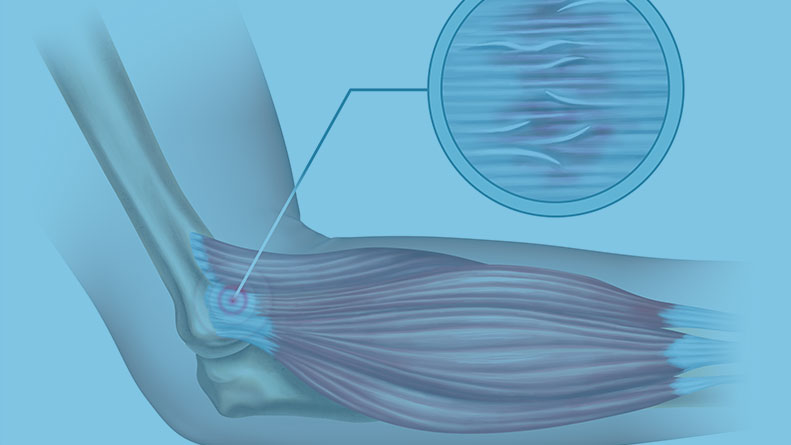
The biceps muscle is present on the front side of your upper arm and functions to help you bend and rotate your arm. The biceps tendon is a tough band of connective fibrous tissue that attaches your biceps muscle to the bones in your shoulder on one side and the elbow on the other side. Overuse and injury leads to fraying of the biceps tendon and eventual rupture. Dr. Gregory DiFelice specializes in diagnosing and treating biceps tendon ruptures.
Causes of Biceps Tendon Rupture
A biceps tendon rupture can either be partial, where it does not completely tear, or complete, where the biceps tendon completely splits in two and is torn away from the bone. The biceps tendon can tear at the shoulder joint or elbow joint. Biceps tendon ruptures that occur at the shoulder are referred to as proximal biceps tendon ruptures. When it occurs at the elbow, it is referred to as a distal biceps tendon rupture. Common causes of biceps tendon ruptures include lowering one’s body during a pull-up or catching a heavy object unexpectedly.
Symptoms of Biceps Tendon Rupture
Symptoms related to biceps tendon rupture include pain in the front of the shoulder or by the elbow. Patients often report a sudden popping sensation accompanied by pain and bruising. It may become difficulty to bend your elbow or raise your shoulder. You may notice the development of a bulge down by your elbow, called a “Popeye” deformity.
Diagnosis of Biceps Tendon Rupture
To identify a biceps tendon tear, Dr. Gregory DiFelice will ask about your medical history and perform a physical examination of your shoulder and elbow. Some imaging tests, such as an MRI scan, may be ordered to confirm the diagnosis.
Treatment of Biceps Tendon Rupture
Biceps tendon ruptures can be treated by non-surgical and surgical methods based off the degree of injury. Partial tears to the Biceps will include non-surgical treatment, such as rest, ice, and immobilization with a sling. Physical therapy exercises may be recommended to improve range of motion and strength. If you have a complete tear of the biceps tendon, surgery may be recommended.
Surgical options for a complete tendon rupture include proximal and distal tendon repair. For a proximal biceps tendon repair, Dr. Gregory DiFelice may use a combination of both arthroscopic and open techniques to reattach the tendon to your humerus (arm bone). For a distal biceps tendon repair, Dr. Gregory DiFelice will make a small incision near the crease of your elbow. The end of the torn tendon will then be stitched and reattached to the bone with the use of a small button. For either procedure, you will be placed into a post-operative sling. Following a biceps tendon repair a rehabilitation program will be started that helps you resume a wider range of activities. Usually, a complete recovery may take about 4 to 8 months depending on your injury pattern.
For more information, please contact our office to schedule your appointment today.
Hamstring Tendon Rupture
What is a Hamstring Tendon Rupture?
The hamstring are comprised of four muscles located on the back your thigh. This grouping includes the semitendinosus, semimembranosus, and the long and short head of the biceps femoris. They are assist in bending your knee and extending your hip. The hamstring tendons are made of tough, fibrous tissue that connects the muscles to both your pelvis and shin bone. Overuse and injury leads to fraying of the hamstring tendons and eventual rupture. Dr. Gregory DiFelice specializes in diagnosing and treating hamstring tendon ruptures.
Causes of Hamstring Tendon Rupture
A hamstring tendon rupture can either be partial, where it does not completely tear, or complete, where the hamstring tendon completely splits away from the bone. The hamstring tendon most completely tears closer to your pelvis, or proximally. These injuries often occur from a sudden forceful contraction of your hamstring muscles, like when you start to run or jump, or from overstretching the muscle.
Symptoms of Hamstring Tendon Rupture
Symptoms related to hamstring tendon ruptures include pain along the back of your thigh. Patients often report a sudden popping sensation accompanied by pain and bruising. It may become difficulty to bend your knee or your lift your leg. You may notice difficulty while walking.
Diagnosis of Hamstring Tendon Rupture
To identify a hamstring tear, Dr. Gregory DiFelice will ask about your medical history and perform a physical examination of your knee and hip. Some imaging tests, such as an MRI scan, may be ordered to confirm the diagnosis.
Treatment of Hamstring Tendon Rupture
Hamstring tendon ruptures can be treated by non-surgical and surgical methods based off the degree of injury. Partial tears to the hamstring will include non-surgical treatment, such as rest, ice, elevation, and compression. Physical therapy exercises may be recommended to improve range of motion and strength. The uses of crutches and protected weight bearing is often used for the first few weeks. The healing time for non-surgical hamstring injuries is typically 6 weeks depending on the injury pattern. If you have a complete tear of the hamstring tendon, surgery may be recommended.
Surgical options for a complete tendon rupture include a proximal hamstring repair. Dr. Gregory will make a small incision along your gluteal crease. The tendons will then be stitched back to your pelvis and anchored into the bone. After, you will be placed into a post-operative brace and asked to walk with crutches for the first few weeks. Following a hamstring tendon repair a rehabilitation program will be started that helps you resume a wider range of activities. Usually, a complete recovery may take about 6 to 9 months depending on your injury pattern.
For more information, please contact our office to schedule your appointment today.
Patellar Tendon Rupture
What is A Patellar Tendon Rupture?
Patellar tendon rupture is the rupture of the tendon that connects the patella (kneecap) to the top portion of the tibia (shinbone). The patellar tendon works together with the quadriceps muscle and the quadriceps tendon to allow your knee to straighten out. Overloading this tendon can result in tearing of the fibers, resulting in instability. Dr. Gregory DiFelice specializes in diagnosing and treating patellar tendon tears.
Causes of Patellar Tendon Rupture
Patellar tendon tear most commonly occurs in people who participate in sports that involve jumping and running. Patellar tendon can be ruptured due to several reasons such as by a fall, direct blow to the knee, or landing on the foot awkwardly from a jump. Chronic inflammation, or tendinosis, can result in micro tearing of the tendon, leading to complete ruptures. The use of medications such as oral steroids and some antibiotics may also weakens the patellar tendon.
Symptoms of Patellar Tendon Rupture
When the patellar tendon tears, the knee cap may lose its anchoring support to the tibia, as a result, when the quadriceps muscle contracts, the knee cap may move up into the thigh. You are unable to straighten your knee and upon standing, the knee buckles upon itself. You may notice a pop with immediate pain and swelling. Some patients report extensive bruising, that may track down to your ankle. Bending you knee may become quite difficult and painful. Additionally, you may notice difficulty with walking.
Diagnosis of Patellar Tendon Rupture
To identify a patellar tendon tear, Dr. Gregory DiFelice will ask about your medical history and perform a physical examination of your knee. Some imaging tests, such as an X-ray or MRI scans may be ordered to confirm the diagnosis. An X-ray of the knee is taken to know the position of the kneecap and MRI scans are used to know the extent and location of the tear.
Treatment of Patellar Tendon Rupture
Depending on the degree of the tear, treatment options may include non-surgical and surgical treatment. Non-surgical treatment consists of rest, ice, compression, and elevation (RICE protocol); all assist in controlling pain and swelling. Physical therapy may be recommended to improve knee motion and strength. A knee brace may be needed to help immobilize your knee. Crutches may be recommended to protect your knee.
Surgical treatment is recommended for patients who have a complete rupture of the tendon. For this procedure, Dr. DiFelice will make an incision along the front of your knee cap. The torn end of the patellar tendon will then be stitched. These stitches will then be passed through your knee cap through small drill holes and tied down, to pull the tendon back into its normal position. You will be placed into a post-operative knee brace and asked to walk with crutches for the first few weeks. Following a patellar tendon repair a rehabilitation program will be started that helps you resume a wider range of activities. Usually, a complete recovery may take about 6 to 12 months depending on your injury pattern.
For more information, please contact our office to schedule your appointment today.
Quadriceps Tendon Rupture
What is A Quadriceps Tendon Rupture?
Quadriceps tendon is a thick tissue located at the top of the kneecap. The quadriceps tendon works together with the quadriceps muscles to allow us to straighten our leg. The quadriceps muscles are the muscles located in front of the thigh. Overloading this tendon can result in tearing of the fibers, resulting in instability. Dr. Gregory DiFelice specializes in diagnosing and treating quadriceps tendon tears.
Causes of Quadriceps Tendon Rupture
Quadriceps tendon rupture most commonly occurs in people who participate in sports that involve jumping and running. Quadriceps tear occurs by a fall, direct blow to the leg, and when you land on your leg awkwardly from a jump. Chronic inflammation, or tendinosis, can result in micro tearing of the tendon, leading to complete ruptures. The use of medications such as oral steroids and some antibiotics may also weakens the quadriceps tendon.
Symptoms of Quadriceps Tendon Rupture
When the quadriceps tendon tears, the knee cap may lose its anchoring support in the thigh as a result the patella moves towards the foot. You will be unable to straighten your knee and upon standing the knee buckles upon itself. You may notice a pop with immediate pain and swelling. Some patients report extensive bruising, that may track down to your ankle. Bending you knee may become quite difficult and painful. Additionally, you may notice difficulty with walking.
Diagnosis of Quadriceps Tendon Rupture
To identify a quadriceps tendon tear, Dr. Gregory DiFelice will ask about your medical history and perform a physical examination of your knee. Some imaging tests, such as an X-ray or MRI scans may be ordered to confirm the diagnosis. An X-ray of the knee is taken to know the position of the kneecap and MRI scans are used to know the extent and location of the tear.
Treatment of Quadriceps Tendon Rupture
Depending on the degree of the tear, treatment options may include non-surgical and surgical treatment. Non-surgical treatment consists of rest, ice, compression, and elevation (RICE protocol); all assist in controlling pain and swelling. Physical therapy may be recommended to improve knee motion and strength. A knee brace may be needed to help immobilize your knee. Crutches may be recommended to protect your knee.
Surgical treatment is recommended for patients who have a complete rupture of the tendon. For this procedure, Dr. DiFelice will make an incision along the front of your knee cap. The torn end of the quadriceps tendon will then be stitched. These stitches will then be passed through your knee cap through small drill holes and tied down, to pull the tendon back into its normal position. You will be placed into a post-operative knee brace and asked to walk with crutches for the first few weeks. Following a quadriceps tendon repair a rehabilitation program will be started that helps you resume a wider range of activities. Usually, a complete recovery may take about 6 to 12 months depending on your injury pattern.
For more information, please contact our office to schedule your appointment today.